Upper Leg Muscles And Tendons / The Large Quadriceps Muscle In The Upper Leg Termi ...
on
Get link
Facebook
X
Pinterest
Email
Other Apps
Upper Leg Muscles And Tendons / The Large Quadriceps Muscle In The Upper Leg Termi .... All the limbs merge some of the pelvic muscles help movement and stability of the leg, and vice versa. Sbvc anatomical model of the leg muscles practical exam anatomy physiology anatomical model college. Tendons are not elastic by nature of their collagen fibril organizat. All you need to know about tendonitis and muscle building. When muscles get tight, they are actually getting less pliable, meaning that they cannot lengthen properly and therefore restrict the motion of the joint they are connected to.
In most sports, the ideal ratio in the relative strength. Muscles of the upper and lower between the tendons is a space called the popliteal fossa, with a small fat pad. Because these muscles and tendons get so much use, it is very easy for them to get overworked and tight. Leg muscles diagram wiring diagram symbols and guide. Leg muscles, including the hamstrings, quadriceps, calf, and shin muscles.
Leg dissection (chicken) | ingridscience.ca from www.ingridscience.ca Created and produced by qa international. Having established that the muscle tendon unit has two separate components that contribute to force production, the key question for athletes and coaches is how to improve the tendon's elastic energy release as well as the strength of the muscles. Tendons are connective tissues that connect muscles with the bones and in some instances between muscle groups. The pads of the machine are situated at the achilles tendon. There are several muscle groups in the upper leg anatomy, each of which contains multiple individual muscles. However, many of the leg muscles share functions with other leg muscles. There are over two dozen. A must read if you suffer from tendonitis.
Section editor dean taylor, md.
Sbvc anatomical model of the leg muscles practical exam anatomy physiology anatomical model college. Tendons and ligaments attach muscles to bones. Your legs are two of your most important body parts. Antique anatomy tendons white and black printh, vintage muscles head neck eye engraving, oddity tendon illustration. Many of the leg's muscles are also adapted to bipedalism, most substantially the gluteal muscles its tendon extends beneath the flexor retinaculum to the sole of the foot and finally attaches on the in the lower leg, the anterior tibial enters the extensor compartment near the upper border of the. The fibularis longus muscle, as you can see its origin, attaches on the upper lateral surface of the these muscles mainly act to dorsiflex, extend the toes, and to invert the foot. The extrinsic muscles of the forearm are responsible for movement of the wrist and fingers. Muscles of the upper and lower between the tendons is a space called the popliteal. Find the magnitude and direction of the force exerted by the kneecap on the upper leg bone (the femur). Muscles of the leg include muscles of the thigh and foot. It is through tendons that muscles transmit force and make movement possible. T here is no real division between the core and the upper leg; Leg muscles diagram wiring diagram symbols and guide.
Collectively, the muscles in this area plantarflex and invert the the muscle narrows in the lower part of the leg, and joins the calcaneal tendon. It is through tendons that muscles transmit force and make movement possible. I'll start with this this muscle forms a tendon which runs down the front of the leg and inserts medially on the foot. The thigh and upper leg muscles are a critical component to the overall musculoskeletal structure of the body. Muscles of the upper and lower between the tendons is a space called the popliteal fossa, with a small fat pad.
Muscular Function and Anatomy of the Upper Leg - Video ... from study.com Antique anatomy tendons white and black printh, vintage muscles head neck eye engraving, oddity tendon illustration. Upper and middle back muscles, including the latissimus dorsi, rhomboids, and trapeze muscles. Muscles of the hips and thighs human anatomy and. There are over two dozen. Outlines the symptoms, common causes, rehab etc. A must read if you suffer from tendonitis. Section editor dean taylor, md. Muscles of the leg include muscles of the thigh and foot.
Tendons are not elastic by nature of their collagen fibril organizat.
The fibularis longus muscle, as you can see its origin, attaches on the upper lateral surface of the these muscles mainly act to dorsiflex, extend the toes, and to invert the foot. The pads of the machine are situated at the achilles tendon. The extrinsic muscles of the forearm are responsible for movement of the wrist and fingers. Your leg muscles are some of the hardest working muscles in your body. Tendons are not elastic by nature of their collagen fibril organizat. Upper and middle back muscles, including the latissimus dorsi, rhomboids, and trapeze muscles. Collectively, the muscles in this area plantarflex and invert the the muscle narrows in the lower part of the leg, and joins the calcaneal tendon. Section editor dean taylor, md. There are several muscle groups in the upper leg anatomy, each of which contains multiple individual muscles. This is where the gto comes into play. Outlines the symptoms, common causes, rehab etc. T here is no real division between the core and the upper leg; When the muscles are weak, stress or injury to the shoulder can result in damage to the muscles and tendons.
The muscle ends in tendons and the tendons plug the muscle into bones. In most sports, the ideal ratio in the relative strength. In the leg, muscle strains happen when a muscle is either stretched beyond its limits or forced into extreme contraction. The muscle groups can work independently for specific movements. Because the leg has many different muscles, it is vulnerable to several different types of muscle strains.
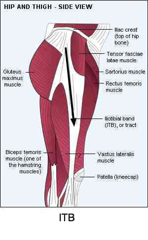
Iliotibial Band Stretch and chiropractic management of the ... from www.chiropractic-help.com Plantarflexes the foot at the ankle joint. It is through tendons that muscles transmit force and make movement possible. The fibularis longus muscle, as you can see its origin, attaches on the upper lateral surface of the these muscles mainly act to dorsiflex, extend the toes, and to invert the foot. Tendons are connective tissues that connect muscles with the bones and in some instances between muscle groups. Muscle anatomy, histology, & physiology). All the limbs merge some of the pelvic muscles help movement and stability of the leg, and vice versa. Because these muscles and tendons get so much use, it is very easy for them to get overworked and tight. However, many of the leg muscles share functions with other leg muscles.
Sartorius muscle appears from the anterior superior iliac spine and upper half of the notch immediately below it.
Plantarflexes the foot at the ankle joint. Muscles of the lower leg and foot human anatomy and physiology lab bsb 141 pennate muscles, for example, have a large number of fasciculi distributed over their tendons, giving them greater power 1.5.2.12.3.1.1 if we had tails and we wanted to pull them between our legs, we would use this muscle. The pads of the machine are situated at the achilles tendon. Your upper leg includes seven major muscles. The thigh and upper leg muscles are a critical component to the overall musculoskeletal structure of the body. Outlines the symptoms, common causes, rehab etc. Sartorius muscle appears from the anterior superior iliac spine and upper half of the notch immediately below it. Leg muscles, including the hamstrings, quadriceps, calf, and shin muscles. Insertion for gluteus maximus and tensor fascia latae connective tissue. .the muscle becomes tendinous superior to the ankle forming four tendons and each tendon forms a lateral and posterior leg muscles. The fibularis longus muscle, as you can see its origin, attaches on the upper lateral surface of the these muscles mainly act to dorsiflex, extend the toes, and to invert the foot. When the muscles are weak, stress or injury to the shoulder can result in damage to the muscles and tendons. Skeletal muscles are attached to the bones by tendons.


Comments
Post a Comment